Microscope is an optical instrument used to magnify minute objects or micro-organism which cannot be seen by naked eye.
In general, Microscope is used in microbiology for two basic purposes:
· The initial detection of microbes and
· The preliminary or definitive identification of microbes.
Microscopic methods:
1. Light microscopy
2. Electron microscopy
3. Scanning probe microscopy
1. Light microscopy
**A light microscope refers to the used of any kinds of microscope that uses natural or artificial transmitted light as the source of light to observe specimens.
In light microscopy, light typically passes through a specimen and then through a series of magnifying lenses. Here microscopy resolving power is an important component.**
Different types of Light Microscopy include
· Bright-field Microscopy
· Dark-field Microscopy
· Dark-ground Microscopy
· Phase-contrast Microscopy
· Interference Microscopy
· Fluorescence Microscopy
A. Bright-field Microscopy:
Bright field Microscopy always refers to as ordinary Light Microscope is the most common form of Light Microscopy that uses a compound Light Microscope.
A compound light microscope primarily consists of a compound lens system that contains a number of objective lens.
With this microscope, specimens are rendered visible
because of the differences in contrast between them and
the surrounding medium.
because of the differences in contrast between them and
the surrounding medium.
Many bacteria are difficult to see well because of their lack of contrast with the surrounding medium
It may be used to examine either wel-films or “hanging drop” for demonstration of the motility of the flagellated bacteria and protozoa. It is also useful for demonstration of the structural details.
B. Dark-ground (Dark-field) Microscopy:
The Dark ground microscopy uses of Dark Ground Microscope, a special type of compound Light Microscope.
This Microscope uses reflected light instead of transmitted light used in the ordinary Light Microscope. It prevents light from falling directly on the objective lens with the result that the micro-organisms appear brightly stained against a dark background. It is useful for very thin bacteria (such as spirochetes) not visible under ordinary illumination, since the reflection of Light Microscope makes them appear larges.
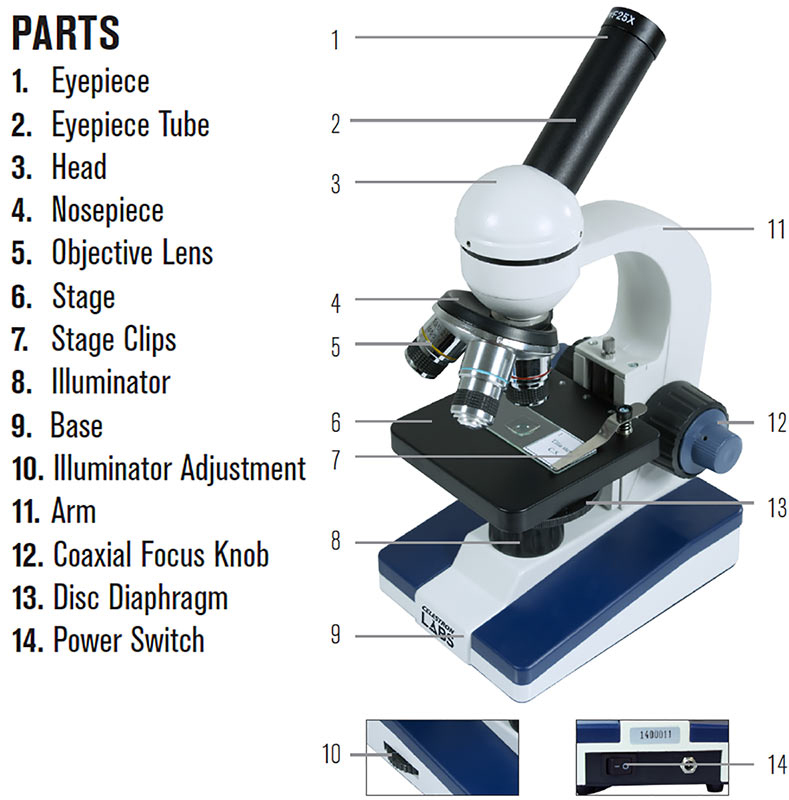 |
| A Compound Light Microscope |
It is also helpful for demonstration of motility of flagellated bacteria and protozoa.
This technique of Microscopy is particularly valuable for observing organisms such as Treponema pallidum, a spirochete which is less than 0.2 mm in diameter and therefore cannot be observed with direct light.
C. Phase-contrast Microscopy:
The Phase-contrast Microscope was developed to improve contrast differences between cells and the surrounding medium, making it possible to see living cells without staining them.
Phase-contrast Microscopy is based on the wave nature of light rays, and the fact that the light rays can “in phase” (their peaks and valleys match) or “out of the phase”.
Phase-contrast Microscopy improves the contrast; the internal structures of a cell become more sharply defined and make evident the structure with in cells that differ in thickness or refractive index. It is used for studying unstained cells, detailed examination of internal structures in living micro-organisms.
*It is used to study Amoeba and Trichomons.
D. Fluorescent microscopy:
Fluorescent is based on the principle that the specimens stained with “fluorescent dye” when exposed to ultraviolet light result in emission of longer wavelength of light.
Fluorescence needs a “Fluorescence Microscope” fitted with an ultraviolet source.
It is used for “direct demonstration” of antigen of a pathogen in clinical specimens by direct “fluorescent antibody test” and also used for estimation of antibodies in the serum by direct fluorescent antibody test.
Fluorescence Microscopy is widely used in clinical diagnostic microbiology. For example, the fluorochrome auramine O, which glows yellow when exposed to ultraviolet light, is strongly absorbed by Mycobacterium tuberculosis, the bacterium that causes tuberculosis. When the dye is applied to a specimen suspected of containing M tuberculosis and exposed to ultraviolet light, the bacterium can be detected by the appearance of bright yellow organisms against a dark background.
E. Interference Microscopy/Confocal Microscopy:
This is another specialized application of Light Microscopy used for demonstrating cell organelles.
*[In Confocal Scanning Laser
Microscopy, a laser beam is bounced off a mirror that directs the
beam through a scanning device. Ten the laser beam is directed
through a pinhole that precisely adjusts the plane of focus of the
beam to a given vertical layer within the specimen.
Microscopy, a laser beam is bounced off a mirror that directs the
beam through a scanning device. Ten the laser beam is directed
through a pinhole that precisely adjusts the plane of focus of the
beam to a given vertical layer within the specimen.
By precisely illuminating only a single plane of the specimen, illumination
intensity drops off rapidly above and below the plane of focus,
and stray light from other planes of focus are minimized.
intensity drops off rapidly above and below the plane of focus,
and stray light from other planes of focus are minimized.
Thus, in a relatively thick specimen, various layers can be observed by
adjusting the plane of focus of the laser beam.]*
adjusting the plane of focus of the laser beam.]*
Its main use is:
1. To obtain three dimensional images of entire cell; by monitoring the distributions and concentrations of substances such as ATP and calcium ions.
2. To evaluate cellular physiology.
Thus, images obtained from different layers can be stored and then digitally overlaid to reconstruct a three-dimensional image of the entire specimen
..
3. Electron Microscopy
The high resolving power of Electron Microscopes has
enabled scientists to observe the detailed structures of prokaryotic and eukaryotic cells.
enabled scientists to observe the detailed structures of prokaryotic and eukaryotic cells.
Electron microscopy utilizes a beam of electrons instead of a bean of light used in the Light Microscope. Rather than using glass lenses, visible light and the eye to observe the specimen, the Electron Microscope uses *electromagnetic lenses*, and a fluorescent screen to produce the magnified image.
There are mainly two types of Electron Microscope in general use:
1. Transmission Electron Microscope (TEM):
In Transmission Electron Microscope, electrons like light pass directly through the specimen that has been prepared by thin sectioning freeze fracturing or freeze etching.
*It is used to observe fine details of cell structure.*
2. Scanning Electron Microscope:
The Scanning Electron Microscope scans a beam of electrons back and forth over the surface of the specimen; producing three dimensional views of the surface of whole micro-organisms.
3. Scanning Probe Microscopy
Another Electron Microscopy is Scanning Probe Microscopy. It is also very useful and popular instrument, in the latest modern research projects and thesis.
It uses the measure surface features by moving a sharp probe over the object’s surface.
Scanning probe Microscopes maps the bumps and the valleys of a surface on an atomic scale.
Their resolving power is much greater than the Electron Microscope and the samples do not need special preparation as they do for Electron Microscopy.
Among the new scanned probe Microscopes are:
1. Scanning Tunneling Microscopy (STM): They are used to provide indirectly detailed views of molecules such as DNA.
2. Atomic Force Microscope (AFM): They produce three-dimensional images of the surface of a molecule.
*Interactions between proteins of the bacterium Escherichia coli can
be studied with the atomic force microscope.
be studied with the atomic force microscope.
Now let’s read about the differences between Light Microscope and Electron Microscope;
Difference between Light Microscope and Electron Microscope
Serial no.
|
Point of difference
|
Light microscope
|
Electron microscope
|
1.
|
Definition
|
A Light Microscope also known as optical microscope is an optical instrument used to make objects larger in order to view their details.
|
An Electron Microscope is an optical instrument that uses abeam of electron ns make objects larger for detailed view.
|
2.
|
Using system
|
Simple to use
|
Users require technical skills.
|
3.
|
Substance to view
|
Can view both live and dead specimens
|
Views only dead specimens.
|
4.
|
Condition of surface
|
Poor surface view
|
Good surface view and internal details
|
5.
|
Illumination
|
Uses light rays to illuminate to specimens
|
Uses a beam of electrons to view specimens
|
6.
|
Lenses
|
Lenses are made of glasses
|
Lenses are made of electro-magnets
|
7.
|
Maintenance
|
Cheap to buy and has low maintenance cost
|
Very expensive to buy and maintenance
|
8.
|
Image type
|
Image is colored
|
Image is black and white.
|
9.
|
Resolving power
|
It has low resolving power micrometer to 0.3micrometrer)
|
It ahs high resolving power (0.0001micrometer), about 250 times higher than light microscope.
|
10.
|
Magnification power
|
It has magnification of 500x to 1500x
|
It has magnification to 1.00, 000x to 3, 00,000x.
|
11.
|
Special requirement
|
Vacuum, filament, cooling system and high voltage electricity are required
|
Vacuum, filament, cooling system and high voltage electricity is very essential requirement.
|
12.
|
Identifying way of image
|
Image is seen identified by eyes through ocular lenses.
|
Image is received in zinc sulfate fluorescent screen on photographic plate
|
13.
|
Risk
|
Radiation is risk is absent
|
There is risk of radiation leakage.
|
14.
|
Specimen
|
Specimen is stained with cooled dyes
|
Specimen is coated with heavy metals in order to reflect electrons.
|
No comments:
Post a Comment